Excerpt
INDEX
Chapter 1 – INTRODUCTION..
1.1) Vici Syndrome
1.2) EPG
1.3) Autophagy and endocytosis
1.4) Pathogenesis of Vici Syndrome
1.5) Toll-like protein receptors (TLRs)
Chapter 2 – AIM OF THE WORK...
Chapter 3 – MATERIALS AND METHODS..
3.1) Materials
3.1.1) Patients clinical data
3.1.2) Cell Cultures
3.2) Methods
3.2.1) Isolation of Peripheral Blood Mononuclear Cells (PBMCs)
3.2.2) Generation of a lymphoblastoid cell lines
3.2.3) PBMCs stimulation with TLR2-9 agonists
3.2.4) Fibroblast stimulation with CpG or IL-1β and NF-kBp65 translocation
3.2.5) CpG-FITC internalization in lymphoblastoid cell lines (LCLs)
3.2.6) Immunofluorescence
3.2.7) Transient gene silencing by siRNA
3.2.8) Transient gene transfection by lipofection
3.2.9) Transient gene transfection by electroporation
3.2.10) Extraction of genomic DNA
3.2.11) Extraction and quantification of the RNA
3.2.12) Reverse transcription and amplification (RT-PCR)
3.2.13) Electrophoresis, extraction from the agarose gel and sequencing of PCR products
3.2.14) Amplification and quantification by real-time PCR
3.2.15) Protein extraction and quantification
3.2.16) Electrophoresis of proteins by SDS-polyacrylamide gel
3.2.17) Western blotting
3.2.18) Plasmid constructs and transformation of bacterial cells
3.2.19) Preparation of plasmid DNA
3.2.20) DQ-BSA internalization
Chapter 4 – RESULTS...
4.1) Molecular genetic analysis
4.2) Analysis of the effects of mutations on the transcribed EPG5
4.3) Analysis of the effects of the mutations on protein EPG5
4.4) Stimulation of PBMC in vitro with TLR2-9 agonists
4.5) Detection of NF-kB p65
4.6) Analysis of intracellular transport of DQ-BSA
4.7) Analysis of intracellular transport of CpG (EAA1, LAMP1, LAMP2)
4.8) Internalization of CpG-FITC in the patient’s and control fibroblasts
4.9) Transient silencing of EPG5 and internalization of CpG-FITC
4.10) Cellular localization of EPG5 in HEK293T cell lines
Chapter 5 – DISCUSSION
BIBLIOGRAFY
Chapter 1 Introduction
1.1) Vici Syndrome
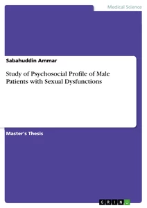
Vici syndrome (OMIM # 242840) is a rare genetic multisystem disorder characterized by five main clinical signs: 1) Agenesis of the corpus callosum (ACC) (total or partial lack of the truncus or “body” of the corpus callosum, connecting the right and left hemisphere of the brain); 2) Cataract (progressive clouding of the lens inside the eye which lead to a decrease in vision); 3) cardiomyopathy(cardiac changes pronounced in the left compared to the right ventricle); 4) Immunodeficiency (functional deficiency of the immune mechanisms); 5) hypopigmentation (variable from total albinism in a partial pigmentation of the retina). (Figure 1)
illustration not visible in this excerpt
Figure 1. Clinical Features of patients with Vici Syndrome. The magnetic resonance brain imaging showed agenesis of the corpus callosum, while examination of the ocular fundus shows a strongly hypopigmental retina. Patients may present dysmorphic facial features, such as microcephaly, hypertelorism, micrognathia.
Vici syndrome is a multisystem disorder that is transmitted as an autosomal recessive trait. The prevalence is unknown at the time and only 20 cases have been reported worldwide (Cullup et al, 2014).
History of Vici Syndrome
In 1988, Dionici Vici et al. described for the first time the clinical case of two brothers with a syndrome of malformations with agenesis of the corpus callosum, cutaneous hypopigmentation, bilateral cataracts, cleft lip (cheiloschisis), cleft palate (palatoschisis), thymus hypoplasia and combined immunodeficiency (Dionisi Vici et al., 1988). The clinical history of the two patients was characterized by severe psychomotor retardation, epilepsy, recurrent infections of the respiratory tract, and chronic mucocutaneous candidiasis. The immunological analysis showed a severe lack of T4+ lymphocytes and IgG2 serum immunoglobulin deficiency. The two brothers died of bronchopneumonia at the age of 2 and 3 years. The clinical and histopathological features seemed to indicate a defect in the organization of the embryonic central nervous system (agenesis of the corpus callosum, hypoplasia of the cerebellar vermis), and of the immune system.
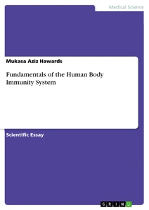
About 10 years later, del Campo et al. described 4 patients (including 2 sibs, a male and a female) with clinical features very similar to those reported by Dionisi Vici. The patients had also agenesis of the corpus callosum associated with oculocutaneous albinism, repeated infections suggestive of an immunodeficiency, cardiomyopathy, growth retardation, postnatal microcephaly and profound developmental delay. Additional central nervous system anomalies present in at least one patient included hypoplasia of the cerebellar vermis, white matter neuronal heterotopia, enlarged extradural space and bilateral schizencephaly (del Campo et al., 1999). The combination of eye findings in these patients was quite specific, including ocular albinism, cataracts, and clinical signs of severely impaired vision. The patients had diffuse retinal hypopigmentation, a finding consistent with ocular albinism. Nystagmus and photophobia could be related to the presence of ocular albinism and CNS abnormalities. Continuous viral, bacterial and fungal infections suggested the clinical case of primary immunodeficiency. The authors hypothesized the existence of a new syndrome, hereafter referred as “Vici syndrome”, probably transmitted as an autosomal recessive trait, because the affected individuals included two brothers of both sexes born to unaffected parents. (Figure 2.)
illustration not visible in this excerpt
Figure 2. Schematic diagram of autosomal recessive inheritance.
Source: http://www.newbornscreening.info/Parents/Images/Autosomal_R_I1.gif
The hypothesis of autosomal recessive inheritance of Vici syndrome was strengthened in 2002 with the clinical description of two new cases, one brother and one sister, by Chiyonobu et al. (Chiyonobu et al., 2002). The two brothers born by healthy parents and showed agenesis of the corpus callosum associated with oculocutaneous albinism, recurrent infections (cutaneous candidiasis, Pseudomonas pneumonia and sepsis), congenital cataracts and cardiomyopathy (markedly and mild dilated left ventricles with extremely reduced contractility, the presence of a muscular bridge between the papillary muscles of the mitral valve, endocardial fibroelastosis).
In the following years, similar clinical cases have been reported that have been contributed to a better definition of the Vici syndrome and a better description of the various clinical features associated with this disease. For example, it has also been reported a neuromuscular involvement in patients suffering from this syndrome (Al-Owain et al.,2010; McClelland et al., 2010; Said et al., 2012). Electro physiologic studies showed severely reduced compound muscle action potentials and a myopathic pattern with pseudomyotonic discharges. Muscle biopsies performed on some of the children, revealed several myopathy features, such as the size of the muscle fibers, central nuclei, enlarged mitochondria and an abnormal accumulation of glycogen. The electron microscope images showed a redundancy of the basal lamina with material between the layers, hypothesizing an exocytosis process of cellular debris.
The renal tubular acidosis is another clinical complication described in only one case report of two brothers with Vici syndrome. In these cases, renal tubular acidosis seemed to occur at the advanced stage of the disease and likely accompanied by cardiomyopathy (Miyata et al., 2007).
The immunological phenotype of these patients is extremely variable, going by a combined immunodeficiency (affecting both T and B cells) to almost normal immune phenotype. The immunological characteristics studied in detail only in some cases (Finocchi et al., 2011; Fennel et al., 2012; Said et al., 2012).
Finocchi et al. described the presence of a heterogeneous immunological phenotype of a patient suffering with Vici syndrome, with respiratory distress and sepsis of unknown origin. The patient revealed a combined immunodeficiency with a productive reduction of absolute lymphocyte count and a reduction of naïve T subsets (CD4+CD45+RA+; CD8+CD45+RA+). In vitro stimulation of peripheral blood mononuclear cells (PBMC) with mitogens and antigen as well as the analysis of peripheral blood TCR repertoire did not reveal any abnormal finding. Serum immunoglobulin and IgG subclasses were normal for age, but he showed unprotective antibody response to tetanus and pneumococcal, with which he been immunized. He had normal absolute count of B cells (CD19+), but memory B cells (CD19+CD27+) were almost absent and peripheral B lymphocytes did not respond to stimulation with CpG. At 2012, Fennel et al. described a patient suffering with Vici syndrome with recurrent infections and progressive leucopenia. The patient also showed a normal mitogenic response of the T cells, humoral response defects with a low number of immunoglobulins and abnormal response of the antibodies. In fact, treatment with intravenous immunoglobulins (IVIG) resulted in a significant improvement of the clinical condition and the disappearance of infectious phenomena Finocchi et al., 2011 and Fennel et al., 2012). Said et al. instead described a child with Vici syndrome, granulocytopenia (corrected spontaneously), with an extremely low number of total lymphocytes and T cells, but normal number of immunoglobulins (Said et al., 2012).
Recently, Cullup et al. analyzed 18 individuals (children from 13 families) with Vici syndrome of European (n=11), Arab (n=3), Turkish (n=2), Japanese (n=1) and British-Asian origin (n=1), 9 of whom were previously reported. Muscle biopsies available from 8 affected individuals showed consistent myopathy features, comprising fiber-type disproportion type 1 atrophy, increase internal nuclei and abnormal glycogen accumulation, as previously reported by Said et al. There were also numerous vacuole-like areas and dense bodies, possibly of lysosomal origin. Myofibrils were lacking in many fibers. Mitochondria were of variable size and showed abnormal distribution and morphology. The condition was severe, with only half of the patients still alive at the time of the study. The most common causes of death were progressive cardiac failure and recurrent infection.
At 2014, Filloux et al. carefully evaluated the ophthalmologic features of Vici syndrome.
Through an exome sequencing analysis, it was possible to identify the gene or -at least one of the genes- responsible for the disease, EPG5 (NM_020964.2); previously KIAA1632) on chromosome 18q12.3 in which there were found mutations in all affected individuals. Analysis of all EPG5 coding exons in 15 families with Vici syndrome with 18 affected individuals showed homozygosity or compound heterozygosity for truncating mutations (including mutations affecting the invariant splice donor and acceptor recognition sites) in 10 individuals and compound heterozygosity for truncating and missense mutations in 2 families.
Diagnosis of Vici syndrome
Until 2013, the diagnosis of Vici syndrome could be carried out based only on the presence of a combination of clinical features (at least 4 of the 5 major clinical signs). The clinical phenotype is variable, but the main clinical signs are usually present at the time of birth, or evolve over time, and the diagnosis is made early in life (Table 1.). The first visible signs after birth are the difficulty in swallowing / feeding and the presence of hypopigmentation on hair and eyes. The MRI shows agenesis of the corpus callosum and the ocular examination may show different ocular abnormalities (bilateral cataract is the most common, but can also be present optic nerve hypoplasia, nystagmus and photophobia). The electrocardiogram is necessary to monitor heart function, as cardiomyopathy can evolve over time leading to progressive heart failure).
The majority of patients suffer from recurrent infections during childhood (which mainly affect the respiratory, gastrointestinal and urinary system, mucocutaneous candidiasis, conjunctivitis and sepsis due to combined immunodeficiency. A skeletal muscle myopathy associated with hypotonia and delayed motor development is frequently found in patients. In some cases, there may be also dysmorphic facial features (micrognathia and cleft lip/ palate) and, rarely, other dysmorphic characteristics (such as syndactyly). Sensor neural hearing loss reported in some children. There may be also other structural abnormalities of the CNS and epilepsy. In addition, some have reported the involvement of the thymus, thyroid, kidneys, as well as hematological abnormalities.
Currently, thanks to the identification of the gene responsible for Vici syndrome, the diagnosis can be confirmed by molecular analysis of the gene EPG5 on chromosome 18q12.3.
illustration not visible in this excerpt
Table 1. List of main clinical signs in patients with Vici syndrome. The table considers all the cases described until 2011 (as amended by Rogers et al., 2011)
Vici syndrome enters into the differential diagnosis with other syndromes, such as the Chediak-Higashi syndrome, which shares common features with Vici syndrome such as a degree of albinism, immunological impairment, and neurological involvement. The EEG features in the patients with Vici syndrome, closely resemble the asymmetric and asynchronous EEG patterns of Aicardi syndrome. Bilateral congenital cataracts that are almost uniformly seen in patients with Vici syndrome, they also be seen in Marinesco-SjÖgren syndrome.
Vici syndrome can be treated, now, only with supportive therapies. Artificial feeding through a nasogastric tube in patients who have feeding difficulties may be required. Recurrent infections should be treated with antibiotic therapy. In patients with severe immunodeficiency, immunoglobulin replacement therapy and antimicrobial prophylaxis should be considered. A therapy with anticonvulsants should be initiated in patients with epilepsy. Cardiac function should be monitored regularly. The congenital cataracts can be surgically corrected and acoustic devices can be of help.
The overall prognosis is not so good (the patients die within the first 2-4 years of life, mainly due to cardiac complications or severe infections), but it can be improved if the diagnosis is made earlier and if the cardiac and immunological aspects are treated appropriately.
In cases where the genetic diagnosis of Vici syndrome has not been established, additional laboratory investigations (for example, transferrin isoelectric focusing), genetic testing and a muscle biopsy will often be performed to exclude multisystem disorders with similar features such as primary glycosylation defects, ciliopathies or mitochondrial disorders. Where performed, supportive features on muscle biopsy include light microscopy abnormalities comprising increased fiber size variability, increased internal nuclei, vacuolization, and ultra-structural changes on electron microscopy such as numerous vacuoles and abnormalities of mitochondrial morphology and localization.
The genetic resolution of a clinical diagnosis of Vici syndrome will help to focus multidisciplinary management and follow-up of the patient. In general, genetic confirmation of the diagnosis will result in a more careful assessment
1.2) EPG5 [ectopic P-granules autophagy protein 5 homolog (C.elegans)]
Vici syndrome is caused by mutations in the gene EPG5 (OMIM # 615068), which encodes an important regulator of the autophagy pathway, the ectopic P-granules autophagy protein 5, involved in the formation of lysosomes. EPG5 is the human homolog of the C.elegans epg5 gene. The gene EPG5 has been cloned for the first time by Nagase et al. by sequencing clones obtained from a size-fractionated fetal brain cDNA library, and was initially named KIAA1632 (Nagase et al., 2000).
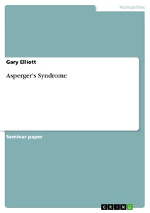
The EPG5 human gene is located on chromosome 18q12.3, has a length of 119,67Kb (NC_000018.10), consists of 44 exons and is transcriptionally driven from the centromere toward the telomere. The messenger RNA (mRNA) is 12633bp long (NM_020964.2) and contains a CDS of 7740 bp translated into a protein sequence of 2579 amino acids (NP_066015.2) with a molecular weight of 280kDa, presumed (Figure 3.). The protein EPG5 is expressed primarily in the central nervous system (CNS), skeletal muscle, heart, thymus, cells of the immune system, lungs and kidneys (Cullup et al., 2013).
illustration not visible in this excerpt
Figure 3. The human gene EPG5. The gene is located on chromosome 18 in the 18q12.3 region, consists of 44 exons coding for a protein of 2579 amino acids.
In a recent work, Cullup et al. analyzed a cohort of 18 individuals with Vici syndrome, identifying mutations in homozygosity or compound heterozygosity in 16 of 18 cases (Cullup et al., 2013). The first EPG5 mutations in the gene were identified by exome sequencing of 4 individuals from 3 families. The other mutations have been found by screening the gene EPG5 in all the remaining patients. All mutations identified so far are truncating mutations (small insertions or deletions) mutations that alter the canonical splice sites. Missense mutations were found in only two cases. Most of the pathogenic variants identified, are private, with only one recurring mutation described (Table 2.). EPG5 mutations are typically inherited from unaffected carrier parents (Cullup et al., 2013; Cullup et al., 2014).
illustration not visible in this excerpt
Table 2. Mutations in EPG5 in patients with Vici syndrome. The sequence variants are described according to the guide line of the nomenclature HGVS (http://www.hgvs.org/) on the sequence GenBank Reference Sequence Number NM_020964.2 (modified by Cullup et al., 2013)
For example in 2 sibs with Vici syndrome, originally reported by Dionisi Vici et al., 1988, Cullup et al., 2013 identified compound heterozygosity for 2 mutations in the EPG5 gene: a 4588C-T transition, resulting in a gln1530-to-ter (Q1530X) substitution, and a 1-bp duplication (5704dupT), resulting in a frame shift and premature truncation. Both patients died by age 3 years.
Among 24 cases of EPG5- related Vici syndrome screened up to date, 38 variants have been detected that have been classed as neutral polymorphisms; identification of heterozygous alleles enables elimination of heterozygous deletions at these loci, but on an individual patient basis, significant proportions of the analyzed region remain uninformative.
illustration not visible in this excerpt
Figure 4. Schematic diagram of EPG-5 gene, which is located on the Crick strand of chromosome 18, showing the location of the missense and truncating mutations in red (Cullup et al., 2013). Missense mutations shown above and truncating mutations below the black bar.
It is important to note that in two individuals (belonging to two different families) have not been found pathogenic variants in the gene EPG5. The analysis of quantitative fluorescent PCR (QF-PCR) showed no change in the number of copies of the gene. The absence of mutations in the gene EPG5 of these individuals does suggest that other genes may be involved (locus heterogeneity), although we cannot exclude the presence of mutations in unanalyzed regions (promoter, regulatory elements, introns).
The gene EPG5 is the human homolog nematode’s Caenorhabditis elegans, epg5, which encodes a protein with a key role in the process of autophagy in multicellular organisms (Tian et al., 2010). Studies performed on muscle biopsies of individuals with mutations in EPG5 showed a severe block in the release of the autophagosomes in the muscles and fibroblasts, resulting in the accumulation of the autophagic cargo in the autophagosomes.
Anomalies in autophagy were found in several diseases, such as certain neurodegenerative diseases, but also in some types of cancer. These findings place the Vici syndrome as a paradigm of human multisystemic disorders associated with defective autophagy and suggest that the autophagic path has a fundamental role in the immune system and in vital organs such as the brain and the heart.
1.3) Autophagy and endocytosis
The autophagy-lysosome system and the ubiquitin-proteasome system are the two major protein degradation systems in eukaryotes, including yeasts, plants, and animals. Autophagy (macroautophagy) was first described as a response to starvation by the liver, with the term “autophagy” derived from the Latin words for “self and “eating”. Later biochemical and morphological studies of autophagy indicated its physiological importance, although its molecular mechanism was unclear because of the absence of molecular markers of autophagosomes and /or autophagy.
To date, at least three types of autophagic pathways have been described, which differ in their routes to lysosomes: macroautophagy (also called simply “autophagy”), microautophagy, and chaperone-mediated autophagy (Figure 5.). Macroautophagy is a degradation system that accompanies the dynamic process of autophagosomes formation, whereas microautophagy involves the sequestering by the lysosome itself of part of the cytoplasm. Both macroautophagy and microautophagy are the nonselective degradation of proteins, lipids, and organelles. In contrast, chaperone-mediated autophagy involves the selective degradation of proteins with a specific-signal sequence called the “KFERQ” motif and is dependent on the molecular chaperone Hsc70. Moreover, chaperone-mediated autophagy does not involve the degradation of lipids or organelles. Lysosome-associated protein-2A functions as a receptor during chaperone-mediated autophagy.
illustration not visible in this excerpt
Figure 5. The three types of autophagy
Source: http://www.nature.com/ncb/journal/v15/n7/images/ncb2788-f1.jpg
About 30 years after the first description of autophagy, autophagic bodies, as intermediate compartments with intraautophagosomal membranes, were first identified in yeast vacuoles under starvation conditions. Taking advantage of yeast genetics, autophagy-defective (atg/apg/aut) mutants of Saccharomyces cerevisiae were isolated as yeasts showing little or no accumulation of autophagic bodies under starvation conditions. The ATG genes contribute to autophagosome formation, with many being well conserved from yeast to mammals (Table 3.).
illustration not visible in this excerpt
Table 3.Autophagy-related genes (National institute of Infectious Diseases)
Source: http://www0.nih.go.jp/niid/biochem/2nd/Slide0014.gif
Autophagy is a process during which cytoplasmic components are degradated in bulk and includes the degradation of proteins, lipids, sugars and organelles (mitochondria, endoplasmic reticulum, and peroxisomes). Extracellular and intracellular signals, including oxidative stress, growth factors, ceramide, ER stress, and glucose, amino acid, and serum starvation can induce autophagy. Autophagy contributes to cellular survival against nutrient deprivation and the turnover of damaged organelles and related to tumorigenesis, neurodegenerative diseases (including Parkinson’s, Alzheimer’s, Huntington’s, and Creutzfeldt-Jakob diseases), cardiomyopathy (e.g., Danon disease), Crohn’s disease, fatty liver, type II diabetes, defenses against intracellular pathogens, antigen presentation, and longevity.
Autophagic flow in mammalian cells: From the formation of isolation membranes to autolysosomes
Following the induction of autophagy, the omegasome is formed from the ER, resulting in the formation of a cup-shaped structure, the isolation membrane, also called the preautophagosome or phagophore. The isolation membrane subsequently elongates to engulf cytoplasmic components, including organelles, and finally closes to form the autophagosomes (initial autophagic vacuole [AVi]), a structure with a double membrane.
Following the formation of the isolation membrane, it elongates to engulf cytoplasmic components. The Atg12-Atg5/Atg16 complex is localized to the cup-shaped structure. In the later stages of isolation membrane elongation, the Atg12-Atg5/Atg16 complex progressively dissociates from the isolation membrane, whereas LC3-II gradually localizes to the both sides of the isolation membrane.
During the process, LC3-I, a cytosolic form of LC3, is progressively lipidated by Atg7, Atg3, and the Atg12-Atg5/Atg16 complex to form LC3-II. Finally, the isolation membrane closes to form the autophagosome (maturation). Although LC3-II is localized to autophagosomes, most of the Atg12-Atg5/Atg16 complex dissociates from the autophagosome. The autophagosome is a double membrane structure, with LC3-II present on both the cytosolic and the intra-autophagosomal surfaces. Therefore, LC3-II increases during autophagosome formation. Rab32 and Rab33 also contribute to the elongation of the isolation membrane. Following autolysosome formation Atg4B delipidates LC3-II on the cytoplasmic surface, to recycle LC3-I (Figure 6.).
illustration not visible in this excerpt
Figure 6. Schematic diagram of autophagy (a) and the LC3 reaction pathway (b)
Source: http://www.nature.com/cdd/journal/v16/n7/images/cdd200954f1.jpg
The outer membrane of the autophagosomes fuses with a lysosome to form an autolysosome (degradative autophagic vacuole [AVd]), a process requiring Rab7. A FYVE and coiled-to-coil domain-containing protein FYCo1 functions as a Rab7 effector, binding to LC3 and PI (3) P, and mediating microtubule plus end-directed vesicle transport. The UVRAG-Vps34-beclin1 PI3-kinase complex and negatively regulated by the Rubicon-UVRAG-Vps34-beclin1 complexes positively regulates the fusion of autophagosomes and lysosomes.
[...]
- Quote paper
- Evangelos Axiotis (Author), 2015, Deciphering the Mechanism of Immune Dysfunction in Vici Syndrome, Munich, GRIN Verlag, https://www.grin.com/document/295415

Similar texts


Publish now - it's free

Comments