Excerpt
Indian mulberry (Morinda citrifolia, Rubiaceae)
Introduction
“Let thy kitchen be thy apothecary; and, Let foods be your medicine.”
Hippocratus
Reducing excess duration of antibiotic therapy is a strategy for limiting the spread of antibiotic resistance, but altering physician practice to accomplish this requires knowledge of the factors that influence physician antibiotic choice (Hassan et al., 2008).
Clinically effective antimicrobial agents exhibit selective nephrotoxicity, with varying degrees, which is characterized by necrosis of the epithelial cells lining the tubules of kidney.
The kidney is especially vulnerable to the hazards of potential drug toxicity because, firstly, it has a rich blood supply, which for a total mass of only about 0.4% of body weight, can amount to 25% of cardiac output.
Secondly, the renal tubular cells may therefore become exposed to drug concentrations which far exceed those encountered in any other body tissue.
Thirdly, the function of the kidney as an obligatory route for the elimination of certain drug means, if extra renal mechanisms of elimination are not activated, drug accumulation will occur.
The higher concentrations of drugs in the circulation make the kidney more susceptible to direct damage, as in the case of nephrotoxicity of certain antimicrobials, especially amino glycosides (Bledsoe et al., 2008).
Aminoglycosides
The aminoglycosidic antibiotics have established their position as highly potent and valuable drugs against infections caused by a wide variety of Gram-negative and Gram-positive bacteria. (Hawkey and Finch, 2007).
Aminoglycosides are structurally composed of aminosugars bound by glycosidic linkages (Kudo et al., 2005) .The number and nature of the aminogroups differeniate each type of aminoglycoside (Carmi Bartal, 2003).
The first and foremost identified and isolated aminoglycoside was Streptomycin from Streptomyces griseus and there are a large number of aminoglycosides available with variations in central ring and its substitutions.
a. Streptomycin – the central ring is streptone and the amino sugar is glucosamine.
b. The central ring is deoxy streptone in neomycin, kanamycin and gentamicin and the aminosugar being pentose and glucosamine for neomycin, kanosamine for kanamycin and garosamine for gentamicin.
c. The central ring is actinamide – spectinomycin .
Aminoglycosides are positively charged in solution, and this cationicity is derived from ionisable aminogroups of the molecule (Holzgrabe et al., 2008). The numbers of ionisable amino groups contained within the molecular structure of the relevant aminoglycosides are six for neomycin, five for gentamicin, tobramycin and netilimicin, four for kanamycin and amikacin and three for streptomycin. Of the members in aminoglycoside group, gentamicin proves to be more effective in its usage on post-operative and infective conditions (Diaz, 2006).
Gentamicin
Structure and Uses
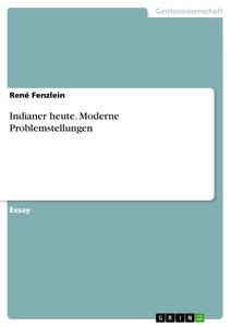
Gentamicin is a broad-spectrum aminoglycosidic antibiotic produced by the actinomycete, Micromonospora purpurea, which was first isolated in 1963. It consists of three major components and numerous minor ones (Fig 1.1)
Gentamicin is one of the few heat-stable antibiotics that remain active even after autoclaving, which makes it particularly useful in the preparation of certain microbiological growth media.
The major components of gentamicin C1, C2 and C1a which differ from each other by the degree of methylation at the 6’ position (Shem-Tov et al., 2003). Chemically gentamicin C1A is 0-3-Deoxy-4-C-methyl-3-(methylamino)-β-Larabinopyranosyl-(1→6)-0-[2,6-diamino-2,3,4,6-tetradeoxy-α-D-erythrohexopyranosyl-(1→4)]-2-deoxy-D-streptamine.
A minor component closely related to the garamicins C’s are C2bA, A1, A2, a3, B, X2, JI-20 A, JI-20B have been well established. Mass spectral analysis of the structure of gentamicin indicates the presence of aminosugar garosamine and 2-deoxystreptamine.
Mode of Action as an Antibacterial
Aminoglycosides like gentamicin "irreversibly" bind to specific 30S-bacterial subunit proteins and 16S rRNA. Specifically, gentamicin binds to four nucleotides of 16S rRNA and a single amino acid of protein S12. This interferes with decoding site in the vicinity of nucleotide 1400 in 16S rRNA of 30S subunit.
illustration not visible in this excerpt
FIGURE 1.1
This region interacts with the wobble base in the anticodon of tRNA. This leads to interference with the initiation complex, misreading of mRNA, with insertion of incorrect amino acids into the polypeptide leading to nonfunctional or toxic peptides and the breakup of polysomes into nonfunctional monosomes.
Human therapeutic dosage of gentamicn against gram-negative bacteria is about 5mg/kg/day, but a dosage above this recommended level and/or constant exposure to the same or high dose, would lead to adverse effect.
Adverse Effects of Gentamicin
Gentamicin, although having established its position as highly potent and valuable drug in the treatment of infections due to a wide variety of gram-negative and gram-positive bacteria, a narrow range exists between its therapeutic to toxic serum concentrations. The clinical application of gentamicin is limited chiefly by the development of nephrotoxicity (Banday et al., 2008). The primary site of aminoglycoside – induced renal injury is at the proximal tubular cells. Nephrotoxicity due to exposure of gentamicin have been well documented in both experimental animals (Chiu et al., 2008) and in clinical trials (Soliman et al., 2007).
Understanding Gentamicin - Induced Nephrotoxicity
The clinical incidence of nephrotoxicity associated with gentamicin is over 10-15%. The primary features of gentamicin nephrotoxicy are functional and morphological alterations in the renal proximal tubule cells, eventually leading to tubular cell necrosis (Juan et al., 2007). Gentamicin induced nephropathy can be studied as a two step process. The first step involves the binding of this drug to the proximal tubular cells (Bledsoe et al., 2008) where they become concentrated and exert their toxic effects. The second step involves the deleterious interaction of these agents with one or more intracellular metabolic processes, which ultimately is expressed as a depression in renal function.
Binding and Uptake of Gentamicin to Proximal Tubular Cells
The interaction between the aminoglycoside - gentamicin and the brush border membrane of the renal proximal convoluted tubules have been described in rats (Hirode et al., 2008) and in rabbits (Hancock et al., 2005). Gentamicin is polybasic due to their side chains containing amino groups and thus is polycationic at physiological pH. Hence the acidic anionic phospholipids of the plasma membrane are prime targets for the charge interaction with the gentamicin (Baronas et al., 2007). Several lines of evidence indicate, particularly phosphoinositides like phosphoinositol (PT), phosphatidylinositol - 4, 5-biphosphate (PI-P2), to be an integral component of the gentamicin binding sites. Of the phosphoinositides, PI-P2 has been shown to have the highest binding affinity for gentamicin followed by PIP and PI (Ogwan et al., 1993). The binding of the drug to the receptor is followed by pinocytosis of the drug-receptor complex with subsequent translocation of the complex to secondary lysosomes. Within the lysosomes, gentamicin might interfere with the catabolism of the receptor by directly inhibiting phospholipase A and C or by modifying substrate – enzyme affinity or by raising the intralysosomal pH above the effective range of the enzyme.
illustration not visible in this excerpt
Inhibition of phospholipase C, results in cortical phospholipidosis, characterized as electron dense phosphatidyl inositol rich-myeloid bodies within lysosomes (Konapska et al., 2007, Dai et al., 2006 and Kizawa et al., 2003). Robert et al. (2007), noted myeloid bodies in the liver and in kidneys of rats following administration of streptomycin and gentamicin. Drug-induced myeloid bodies were also studied by Kaloyanides (1992), suggested that this myeloid body formation within lysosomes, may be due to delay in the degradation toxicity. The second step involves the adverse interaction of gentamicin with one or more critical intracellular processes leading to lysosomal distension and rupture, resulting in functional disruption of the other membrane fluidity. (Fig. 1.2).
Strategies to mitigate gentamicin - induced toxicity
In the hope of preserving antibacterial efficacy while concomitantly preventing or attenuating nephrotoxicity, many studies were designed to elucidate the mechanism of gentamicin-induced renal injury.
Two studies have been conducted
(1) To assess the protein level of a diet that would give the best renal outcome with gentamicin administration, and
(2) To get a better idea about the rhythms of food ingestion associated with the different protein levels.
Therefore, while a protein-rich diet can be protective against gentamicin-induced nephrotoxicity, (Narayana, 2008; Zeeni et al., 2007). Oxidative stress and nitrosative stress are reported to play important role in gentamicin nephrotoxicity (Dhanarajan et al., 2006).
Studies indicate that CoQ10 and green tea due to their antioxidant activity modified the biochemical changes occurred during gentamicin nephrotoxicity and thus had a potential protective effect (Upaganlawar et al., 2006).
A correlation is established between individual features of the lipid peroxidation process and the state of the system of antioxidant protection of the kidney in intact rabbits and their susceptibility to gentamicin nephrotoxicity. The level of gentamicin-induced damage of the kidney is more significant in rabbits with increased concentration of malondialdehyde in the kidney, which is generated by a NADPH-dependent enzyme system. A decreased level of reduced glutathione, as well as the enzymopathy with respect to superoxide dismutase and catalase is among the factors aggravating gentamicin-induced kidney lesion (Bushma et al., 2006).
Administration of a cationic molecule, such as the calcium channel blockers verapamil and nitrendipine, which may compete with the aminoglycoside for binding to anionic membrane lipids leading to its mitigation (Ali et al., 2003).
Administration of polyaspartic acid, a polyanion that binds to the aminoglycoside, forming a nontoxic complex. This binding appears to occur within the acid milieu of the lysosomes in the proximal tubular cells, thus, toxicity, but not renal cortical accumulation of the aminoglycoside is prevented (Kishore et al., 1990).
Administration of sodium bicarbonate to alkalinize the urine, by raising the pH, will decrease the net positive charge on the aminoglycoside thereby diminishing its uptake by the tubular cells (Sassen et al., 2006). Although these findings are intriguing, their applicability in clinical trials remains to be proven.
“Botany and medicine came down the ages hand in hand until the seventeenth century; then both arts became scientific, their ways parted, and no new herbals were compiled. The botanical books ignored the medicinal properties of plants and the medical books contained no plant lore.” Hilda Leyel.
Medicinal plants
Medicinal plants are an important subset of biodiversity which has lately emerged as a hot topic of discussions at various national and international floras. Medicinal plants have a unique significance in India and have remained a part of our cultural ethos from time immemorial. They meet the health needs of large sections of population, provide raw materials to various Indian systems of medicine and are an important source of livelihood to forest dwelling communities. India with its rich resource base and traditional knowledge regarding the use of medicinal plants has the potential to emerge as a world leader in this sector (Latheef et al., 2008; Gold man, 2001).
Herbal medicine
People on all continents have used hundreds of indigenous plants for treatment of ailments since prehistoric times. There are evidence from the Shanidar Cave in Iraq, that suggests, Neanderthals those lived 60,000 years ago used medicinal plants. A body that was unearthed had been buried with eight species of plants which are still widely used in ethnomedicine around the world (Lietava , 1992).
The use of herbs to treat disease is almost universal among non-industrialized societies. A number of traditions came to dominate the practice of herbal medicine at the end of the twentieth century (Xu et al., 2008).
Many of the pharmaceuticals currently available to physicians have a long history of use as herbal remedies, including opium, aspirin, digitalis, and quinine. The World Health Organization (WHO) estimates that 80 percent of the world's population presently uses herbal medicine for some aspect of primary health care (Rossi et al., 2008). Herbal medicine is a major component in all traditional medicine systems and a common element in Ayurvedic, homeopathic, naturopathic, traditional Chinese medicine, and Native American medicine. According to WHO, 74% of 119 modern plant-derived pharmaceutical medicines are used in ways that are similar to their traditional uses.
The use of and search for, drugs and dietary supplements derived from plants have accelerated in recent years. Approximately 25% of modern drugs used in the United States have been derived from plants and approximately 120 active compounds are currently isolated from the higher plants and widely used in modern medicine today and 75 percent show a positive correlation between their modern therapeutic use and the traditional use of the plants from which they are derived (Efferth et al., 2007). More than two thirds of the world's plant species - at least 35,000 of which are estimated to have medicinal value - come from the developing countries and approximately 7,000 medical compounds in the modern pharmacopoeia are derived from plants.
Scenario in India
It has been estimated, that India is endowed with about 47,000 species of plants and ranks 8th in the world biodiversity. Out of these, 8000 species are known to be medicinal. Indian system of Medicine (ISM) use around 2,500 plant species belonging to more than 1,000 genera. About 800 species are used by industry, of which approximately 25% are presently cultivated, (Gold man, 2001).
Anti-nephrotoxic plants
The use of plants for healing by any cultural group is integrally related to local concepts of the nature of disease, the nature of plants, and the world view of the culture. The physical and chemical properties of the plants themselves also bear on their selection by people for medicines, as does the array of plants available for people to choose from (Subramanyam et al., 2008). Other key concepts are that of balance-avoidance of excess, and respect, for self and for others, which are seen as important in living a good life and in maintaining health. As an aspect of respect, attitude is important in collection and use of plants, which are seen as entities with their own intrinsic power to help and heal, or to withhold help (Leslie Main Johnson, 2006).
Valeriana officinalis var. latifolia has the protective effect on lipid-induced nephropathy, and the inhibition of TGF-β 1 expression might be the suggested mechanism of Valeriana officinalis var. latifolia on renal protection (Si et al., 2003).
Besides liver, other organs (lung, kidney and fore stomach) were also influenced favorably by Aloe vera in order to detoxify reactive metabolites, including chemical carcinogens and drugs (Anitha et al., 2006; Singh et al., 2000). Adhatoda, is knowing to increase the potential of the machinery associated with the detoxification of xenobiotic compounds (Singh et al., 2000).
Scope of the Present Investigation
Gentamicin, a member of amino glycoside antibiotics, is polycationic in nature and indeed the most viable broad-spectrum antibiotic. It is active against gram-positive and gram-negative as well as acid fast mycobacterium. Clinical experience associated with use of gentamicin is immense, since it has now entered the fifth decade of their application in the management of patients from the pediatric to the geriatric – age range.
Although the clinical efficacy and superiority of gentamicin is acknowledged when they are used in appropriate clinical settings, nonetheless, these compounds continue to manifest a high profile to undesirable side effects such as nephrotoxicity, ototoxicity and rarely neuromuscular blockade. A narrow therapeutic-to-toxic ratio exists, requiring pharmacokinetic monitoring during administration of gentamicin. It has been estimated that the clinical incidence of gentamicin use is over 10%. The primary features of gentamicin nephrotoxicity are functional and morphological alteractions in the proximal tubular cells, eventually leading to tubular cell necrosis (Zager et al., 2007).
The pathogenesis of gentamicin nephrotoxicity can be viewed as a two-step process. The first step involves the transport and accumulation of antibiotic in high concentration by the proximal tubular cells and is base on the electrostatical binding of the polycationic gentamicin with anionic phospholipids (mainly phosphatidyl inositol, 4,5 – biphosphate). The second step, involves the adverse interaction between these polycationic drugs with one or more critical intracellular processes (Hostetler et al., 1990).
One potential strategy for mitigating the risk of aminoglycoside nephrotoxicity is to inhibit the uptake of drug by the renal proximal tubular cells. Different protocols have been carried out in experimental setting in order to afford protection from renal damage. Successful approaches include dietary manipulation in calcium content and simultaneous administration of substances, able to interfere with gentamicin uptake, such as polyaspartic acid (Guo et al., 2001) and pyridoxal-5-phosphate (Ali et al., 1993).
Recently experiments have been performed to evaluate the effect of different antioxidants on gentamicin – induced renal dysfunction. Medicinal plants have established their position as nephroprotectants in gentamicin induced renal toxicity. Of many medicinal plants, with chemotherapeutic property the most important of it is Morinda citrifolia.
Morinda citrifolia
Kingdom : Plantae
Division : Magnoliophyta
Class : Magnoliopsida
Order : Gentianales
Family : Rubiaceae
Genus : Morinda
Species : Morinda citrifolia
Botanical Name : Morinda citrifolia
Popular Name(s) : Noni, Indian Mulberry, Dibri-Apra, Patje, Mengkudu.
Parts Used : Fruits, Leaves & Roots
Habitat : Grows in shady forests as well as on open rocky or sandy
shores.
Description : It can grow up to 9 m tall, and has large, simple, dark
green, shiny and deeply veined leaves.
Uses : Hawaiian Noni is used for everything like joint pains, immune problems, pain relief, cellular regeneration and more. Noni fruit contains a number of enzymes (proteins) and alkaloids that are believed to play a pivotal role in good health.
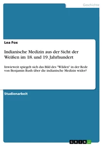
Noni is the Hawaiian name for the fruit of Morinda citrifolia L. (Rubiaceae). Its various vernacular names include: ‘‘Indian mulberry’’, ‘‘nuna’’, or ‘‘ach’’ on the Indian subcontinent, ‘‘mengkudu’’ in Malaysia, ‘‘nhau’’ in Southeast Asia, ‘‘painkiller bush’’ in the Caribbean, or ‘‘cheese fruit’’ in Australia (Morton, 1992; Nelson, 2001; Wang et al., 2002). Noni is native from Southeast Asia to Australia and is cultivated in Polynesia, India, the Caribbean, Central and northern South America (Dixon et al., 1999). The Polynesians have been using the noni plant for food and medicinal purposes for more than 2000 years (Wang et al., 2002). In traditional Pharmacopoeia, the fruit is claimed to prevent and cure several diseases (Issell et al., 2005). It is primarily used to stimulate the immune system and thus to fight bacterial, viral, parasitic and fungal infections; it is also used to prevent the formation and proliferation of tumors, including malignant ones (Wang et al., 2005; Dixon et al., 1999).
illustration not visible in this excerpt
Noni juice is also claimed to relieve inflammation (Xu et al., 2006). Most noni is consumed as juice, although leaves, flowers, bark and roots can also be used (Jensen et al., 2004; McClatchey, 2002; Dixon et al., 1999).
Noni has recently been the object of many claims concerning its nutraceutic properties. Various publications have shown that noni can be used to relieve different diseases, and its registered uses span the Pacific and Asia, as well as Africa. Clinical studies reported a relief of Liver associated with noni consumption (Wang et al., 2002): the observed beneficial effects may result from certain compounds such as scopoletin, nitric oxide, alkaloids and sterols, and also to the antioxidant potential of noni. As a result of this reputation, consumption of this fruit is currently high, not only in the producing countries, but also in the United States, Japan and Europe. In response to this demand, some countries such as Costa Rica and Cambodia have increased the fields being cultivated for noni. In these countries, the fruit is often commercialized fresh or as juice in both formal and informal markets, but it is also found as pasteurized juice, either pure or mixed with other juices (usually grape or blackberry). Commercial interest in noni has tremendously increased in recent years, as provided by the number of patents registered. In the United States, 19 patents have been registered by the US Patent and Trademark Office since 1976 (USPTO, 2005). Noni juice has been recently accepted in the European Union as a novel food (European Commission, Scientific Committee for Food, 2002). Nevertheless, despite the real market opportunities, there has been little scientific research to review the actual nutritional and functional properties of noni products. Furthermore, the phytochemical compounds responsible for their alleged properties have not yet been reviewed.
Considering the non-toxic effects of Morinda citrifolia, the aim of the present study is to establish the protective role of Morinda citrifolia against gentamicin nephrotoxicity and the therapeutic efficacy of Morinda citrifolia.
The present study is focused with the following main objective to access the medicinal properties of Morinda citrifolia on certain cellular metabolism process under the following major headings,
1. Acute toxicity study for determination of LD50.
2. Invitro antioxidant activity of Morinda citrifolia.
EVALUATION OF LD50
INTRODUCTION
The present study aims to investigate the possible toxicity of the Morinda citrifolia and a safety evaluation of the aqueous and methanol extract focusing on its acute toxicity in rats.
In the absence of an efficient primary health care system, traditional medicine occupies a central place in the provision of health care, especially among rural communities of developing countries (Huang et al., 2008). The strong historical bond between plants and human health is well substantiated by plant species diversity and related knowledge of their use as herbal medicines (Tabuti et al., 2003). In addition, it is attributable to the accessibility and affordability of herbal medicines (Steenkamp, 2003).
In order to support, an application for a clinical trial or for the registration of a new drug, it is necessary to satisfy legislation that requires, that certain data should be produced from a variety of toxicological investigations that show the safety profile of the compound to which humans may be exposed. Therefore, in the majority of cases of evaluation of the toxicity of most substances, rodents and non-human primates are first used in preclinical animal safety studies before further studies are done in humans (Sasseville et al., 2008). These animals are mainly used because of their biological similarity to humans that allows them to be regarded as the suitable metabolic models for humans in a broad range of investigations (Pascoe, 1983; Loomis and Hayes, 1996).
An acute toxicity test is a single test that is conducted in a suitable animal species and may be done for essentially all chemicals that are of any biologic interest (Suski et al., 2008). Its purpose is to determine the symptomatology consequent to administration of the compound and to determine the order of lethality of the compound. The test consists of administering the compound to the animals on one occasion (Loomis and Hayes, 1996; Timbrell, 2002).
Furthermore, acute toxicity tests are those designed to determine the effects, which occur within a short period after dosing. They serve to establish the lethal dose range of the test substance and provide prompt warning if a highly toxic compound is being dealt with. They also provide information on the limiting toxicity arising from the pharmacological effects of the compound on target organs and, often, on the maximum dose to be used in subsequent sub-acute studies (chronic studies). This latter information is particularly important for predicting the amount of chemical required for future toxicological studies (Poole and Leslie, 1989). Since a great range of concentrations or doses of various chemicals may be involved in the production of harmful effects, the LD50 has been used by some to devise categories of toxicity on the basis of the amounts of the chemicals necessary to produce harm (Loomis and Hayes, 1996).
MATERIALS AND METHODS
MATERIALS
Animals Used
Male albino rats of Wistar strain weighing between 100-150 gm were obtained from Small Animal Breeding Center, Kerala Agricultural Unviersity, Trissur. The animals were housed in larger spacious cages and they were given food and water ad libitum during the course of the experiment. The animal room was well ventilated and the animal had a 10 ± 1 hour night schedule, through out the experiment period. The temperature remained between 30ºC to 37ºC with only 5ºC differences. Clearance for the handling of experimental animals were obtained from the Ethical Committee CPCSEA No. 659 / 02 / A.
Diet Used
The commercial pelleted animal feed marketed by M/s Hindustan Lever Limited, Bangalore, India, under the trade name of “Gold Mohur rat feed” was used.
Chemicals Used
Sources of Fine Chemicals
All chemicals and solvents used were analytical grade, and purchased from SD Fine Chemicals, Himedia and Qualigens, India.
Plant Material
The fruits of the plant Morinda citrifolia Linn. L. were collected from Coimbatore, Tamilnadu, India in the month of April 2006. A Voucher specimen (No.PP.93558) has been deposited in Botanical Survey of India, Coimbatore, Tamilnadu, India.
Preparation of Extract
The fruits were cut into pieces and shade dried at room temperature. The dried fruits were subjected to size reduction to a coarse powder by using dry grinder and passed through sieve. One kilogram of fruit yielded 160gm of dry material. This powder was packed into soxhlet apparatus and extracted successively with distilled water and 50% methanol (yielded 8 and 8% respectively). The extracts was dried at 45ºC in hot air oven till solid to semisolid mass was obtained and were stored in airtight containers in refrigerator below 10ºC.
Route of administration
Oral administration
The oral route is probably one of the most common means by which a chemical enters the body. Most orally administered chemicals can otherwise have a systemic effect on the organism only after absorption. Oral administration of chemicals that are rapidly absorbed from the gastrointestinal tract would theoretically expose the liver to concentrations of the agent that would not be obtained if other routes of administration were used (Waynforth, 1980; Loomis and Hayes, 1996).
METHODS
The crude fruit extracts of Morinda citrifolia (aqueous and methanol extracts) were tested for its acute toxicity in rats. The study was designed on the basis of OECD (Organization for Economic Co-operation and Development) guideline - 423, adopted on June 2001. An initial test was done to determine the approximate lethal and non-lethal doses of the extracts according to Turner (1965). Five groups of six rats each were used in the experiments. To determine acute toxicity, a single oral administration of the extracts at 4 dose levels (5, 50, 300, 2000mg/kg bw) were tried. The dose levels tested 5, 50, 300, 2000 mg/ml suspensions were prepared in 1% W/V CMC and given at the dose of 1ml / kg bw. The control group were administered an equal volume of water. After administration of the extracts, the animal were observed individually for 4 hrs and there after for 14 days. The parameters noted were grooming, hyperactive, sedation, loss of righting reflex, respiratory rate and convulsion. All animals were observed twice daily for mortality during the period of study (Ghosh, 1984).
RESULTS AND DISCUSSION
Acute toxic studies of Morinda citrifolia (aqueous and methanolic extracts) on experimental rats is depicted in table (2.1).
illustration not visible in this excerpt
In the acute toxicity study, the crude extracts of Morinda citrifolia did not show any significant toxic signs when observed for the parameters during the first 4 hrs and followed by daily observations for 14 days and no mortality was observed. The extract was found to be safe at the tested dose level of 2000 mg/kg bw. The acute toxicity of the extract showed no death of these rats even under high dose levels indicating the high margin of safety of this extracts.
Tahitian Noni juice given daily to rats for 13 weeks at oral doses up to an equivalent of 80 ml/kg body weight caused no signs of toxicity. This indicates that there is a considerable margin between the highest doses administered to rats, which was without any untoward effects (Deng et al., 2007; Glerup, 2001).
SUMMARY AND CONCLUSION
Although, it is the normal practice to determine the LD50, now it is acceptable to limit the study with an acute toxicity test using several doses including reasonably high dose of the extract. The acute toxicity study of Morinda citrifolia (aqueous and methanol extracts) showed no death of these rats even under high dose levels (2000mg/kg bw) indicating the high margin of safety of these extracts.
INVITRO ANTIOXIDANT STUDIES OF Morinda citrifolia
INTRODUCTION
A free radical has been defined as ‘any species capable of independent existence that contains one or more unpaired electrons’. This situation is energetically unstable, making such species often highly reactive and short-lived (Altun et al., 2007). The term ‘reactive oxygen species’ (ROS) and ‘oxidants’ are introduced to describe collectively not only the oxygen derived free radicals, namely super oxide (Oº2) and hydroxyl (OHº), but also the non-radical oxidants like hydrogen peroxide (H2O2) and hypochlorous acid (HOCl) which do not have unpaired electrons. Endogenous production of free radicals occurs continuously during cellular metabolism. Exogenous sources like drugs, pesticides, pollutants, ionizing radiation are known to stimulate the production of free radicals (Facundo et al., 2007).
All cells are known to possess an array of enzymes and compounds (antioxidant defenses) to protect the host from the initiation or propagation of free radical/oxidant damage (Kim et al., 2008; Khan et al., 2007).
Depletion of antioxidant defenses and /or rise in ROS production can tip the ROS-antioxidant balance and cause oxidative stress which may result in tissue injury (Sun et al., 2008). This free radical oxidative stress has been implicated in the pathogenesis of a variety of human diseases (Hamilton et al., 2007).
Oxidative stress can produce major interrelated derangements of cellular metabolism, including peroxidation of lipids (Zabłocka et al., 2008). All cells have developed several lines of defense against this oxidative attack. Their function is to keep free radical under control at physiological levels (Lee et al., 2007). Malondialdehyde (MDA), an end product of lipid peroxidation is increased, suggesting loss of membrane fluidity and transport in the renal cells (De et al., 2007). Many plants are known to posses antioxidant activity (Umukoro et al., 2008; Jeffers et al., 2007), and present study is an attempt to evaluate the antioxidant potency of Morinda citrifolia in an invitro model.
MATERIALS AND METHODS
MATERIALS
The freshly prepared aqueous and methanolic extract of Morinda citrifolia was taken for the present study (50, 500 and 1000 μg/ml).
METHODS
ASSAY OF 1-DIPHENYL 1-2 PICRYLHYDRAZYL (DPPH) RADICAL SCAVENGING ACTIVITY
Principle
The hydrogen atom or electron donation abilities of the corresponding extracts were measured from the bleaching of the purple-coloured methanol solution of 1, 1- Diphenly-2-picrylhydrazyl (DPPH).
Procedure
The scavenging activity of methanol extracts on DPPH radicals was measured according to the method of Shimada et al. (1992). Methanolic solutions of DPPH (10-4) and extract solution were mixed so that the final mass ration were extracts: DPPH=5.5:1 and reference compound: DPPH=0.5:1. The samples were incubated for 15 min in the dark at 30ºC, and the decrease in absorbance at 517 nm was measured against methanol using a spectrophotometer.
ASSAY OF HYDROXYL RADICAL SCAVENGING ACTIVITY
Reagents
1. 1 mM FeCl3
2. 1 mM EDTA
3. 20 mM H2O2
4. 1 mM L-ascorbic acid
5. 30 mM deoxyribose in potassium phosphate buffer (pH 7.4)
6. 2.8% (w/v) trichloroacetic acid
7. 1% (w/w) 2-thiobarbituric acid
Procedure
Hydroxyl radical scavenging activity of extract was measured according to the method of Halliwell et al., (1987). One milliliter of the final reaction solution consisted of aliquots (500 μL) of various concentrations of the extract, 1 mM FeCl3, 1 mM EDTA, 20 mM H2O2, 1 mM L-ascorbic acid and 30 mM deoxyribose in potassium phosphate buffer (pH 7.4). The reaction mixture was incubated for 1 hr at 37ºC, and further heated in a boiling water bath for 15 min after addition of 1 mL of 2.8% (w/v) trichloroacetic acid and 1 mL of 1% (w/w) 2-thiobarbituric acid. The color development was measured of 532 nm against a blank containing phosphate buffer.
ASSAY OF REDUCING POWER ACTIVITY
Reagents
1. Phosphate buffer (200 mM, pH 6.6)
2. 1% potassium ferricyanide
3. 10% tricholoracetic acid
4. 0.1% ferric chloride
Procedure
The reducing power of extracts was determined by the method of Yen and Duh (1993). Different concentration of extracts was mixed with 2.5 mL of phosphate buffer (200 mM, pH 6.6) and 2.5 mL of 1% potassium ferricyanide. The mixtures were incubated for 20 min at 50ºC. After incubation, 2.5 mL of 10% tricholoracetic acid were added to the mixtures, followed by centrifugation at 650xg for 10 min. The upper layer (5 mL) was mixed with 5 mL of distilled water and 1 mL of 0.1% ferric chloride and the absorbance of the resultant solution were measured at 700 nm.
ASSAY OF HYDROGEN PEROXIDE – SCAVENGING ACTIVITY
Reagents
1. 0.1 M phosphate buffer (pH 7.4)
2. 43 mM solution of hydrogen peroxide
Procedure
The extract was dissolved in 3.4 mL of 0.1 M phosphate buffer (pH 7.4) and mixed with 600 μL of 43 mM solution of hydrogen peroxide. The absorbance value (at 230 nm) of the reaction mixture was recorded at 10 min intervals between zero and 40 min. for each concentration, a separate blank sample was used for background subtraction (Ruch et al., 1989).
ASSAY OF FERRIC REDUCING ANTIOXIDANT POWER (FRAP)
Reagents
1. Acetate buffer - 300m M pH 3.6
2. Dilute HCl - 40mM,
3. TPTZ (2,4,6-tripyridyl-s-triazine) - 10mM
4. Ferric Chloride - 20mM.
Standard
Ferrous sulphate – Prepared 3mM solution in 1 litre distilled water.
Procedure
The freshly prepared FRAP reagent comprised of 300 mM acetate buffer, pH 3.6; 10 mM 2,4,6-tripyridyl-s-triazine in 40 mM HCl; and 20 mM FeCl2 at a 10:1:1 ratio. 300 μl of working FRAP reagent was mixed with 10 μl of the extract of Morinda citrifolia or ascorbic acid. The latter was used to calibrate the FRAP assay. The mixtures were incubated for 4 min at 30ºC. The change in absorbance was monitored at 610 nm, and was translated into a FRAP value. Antioxidant activity was calculated and expressed as ascorbic acid equivalent per mg of extract (μg/mg) (Benzie and Stains, 1999).
RESULTS AND DISCUSSION
Antioxidants are molecules which can safely interact with free radicals and terminate the chain reaction before vital molecules are damaged.
A free radical attack on the membrane usually damages a cell to the point that it must be removed by the immune system (Chin et al., 2008). If, free radical formation and attack are not controlled within the muscle during exercise, a large quantity of muscle could easily be damaged (Pierce et al., 2008). Protection from damage occurs through the action of excellent defense system consisting of antioxidants, some endogenously produced and some provided through dietary intake (Flora et al., 2007).
Antioxidants can interfere with the oxidation process by reacting with free radicals, chelating catalytic metals, and also by acting as oxygen scavengers (Narayana, 2008). However, antioxidant supplement may be used to the help the human body reduce oxidative damage (Emara et al., 2008; Zin et al., 2002).
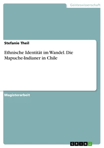
DPPH Radical Scavenging Activity of Morinda citrifolia extract
The DPPH radical is considered to be a model of lipophilic radical (Jimenez-Escrig et al., 2007). A chain reaction in lipophilic radical was initiated by lipid autoxidation. DPPH is a stable free radical and the assay determines the ability of the test sample to reduce the radical to the corresponding hydrazine by converting the unpaired electrons to paired ones (Letelier et al., 2007).
illustration not visible in this excerpt
1, 1-Dipheny-2-picrylhydrazyl (DPPH) is one of the stable nitrogen centered free radical and a free radical representative to be effectively scavenged by antioxidants (Villano et al., 2007). This DPPH assay has been widely used to test the ability of plant extract to act as free radical scavenger (Milioli et al., 2007).
Aqueous and methanolic extracts of Morinda citrifolia showed DPPH radical scavenging activity with 66% and 72% at concentration of 1000 μg, respectively (Table -3.1), but, the effect was relatively lower as compared to that of standard BHT and ascorbic acid. Of all samples tested, methanolic extract showed the highest scavenging activity (28-72% at 50-1000 μg/mL).
illustration not visible in this excerpt
Reducing power activity of Morinda citrifolia
Antioxidant activity has been reported to be concomitant with the development of reducing power (Tanaka et al., 1988). Okuda et al. (1983), reported that the reducing power of tannins prevents liver injury by inhibiting the formation of lipid peroxides. The reducing capacity of a compound may serve as a significant indicator of its potential antioxidant activity (Mier et al., 1995).
illustration not visible in this excerpt
At concentration of 1000 μg/mL, aqueous and methanolic extracts showed absorbance of 0.419 - 0.857 and 0.523 – 1.091, respectively (Table-3.2), thus, suggesting a reducing activity of the plant extract. The reducing power might be due to hydrogen-donating ability (Shimada et al., 1992), and is generally associated with the presence of reductone (Duh, 1998). Since, phenolic compounds present in the extract are good electron donors (Kaur et al., 2008), they may accelerate the conversion of H2O2 to H2O (Ruch et al., 1984).
illustration not visible in this excerpt
Hydroxyl radical scavenging activity of Morinda citrifolia
Hydroxyl radicals are the major active oxygen species causing lipid oxidation and biological damage (Conde de la Rosa et al., 2008; Aurand et al., 1977). The most reactive of the ROS that attack almost every molecule in the body is the hydroxyl radical. It initiates the peroxidation of cell membrane lipids yielding malondialdehydes, (Pialoux et al., 2006) which is considered as mutagenic and carcinogenic. Hydroxyl radicals are formed invivo from water by high-energy irradiation or from H2O2 in a metal-catalyzed process (Domijan et al., 2007).
[...]
- Quote paper
- Anitha Thiyagarajan (Author), 2009, Indian mulberry (Morinda citrifolia, Rubiaceae), Munich, GRIN Verlag, https://www.grin.com/document/201756
Similar texts


Publish now - it's free

Comments